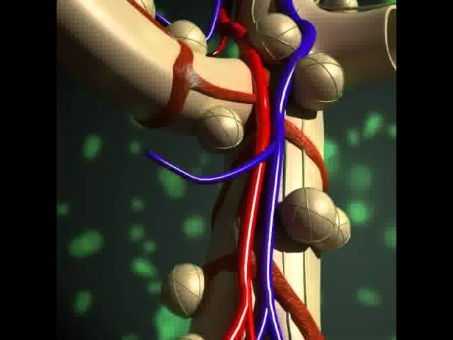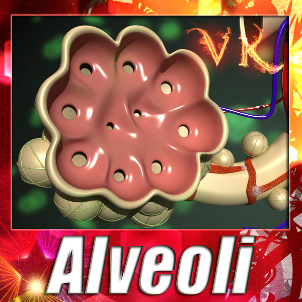


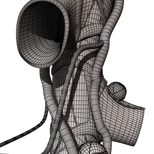
Alveoli Anatomy High resolution textures. Smootheable.
3000 x 3000 high resolution textures.
Unsmoothed model included.
**************************************************************
-Model is centered at 0,0,0.
-All object are named.
-All materials are named.
-No unnecessary objects.
-Model looks like the thumbnails.
**************************************************************
Obj, fbx and max files, are compressed in zip files with all materials, textures and folders.
High resolution textures color, bump and reflect map. Standard 3ds max materials and Vray materials included. with .mat and .mtl
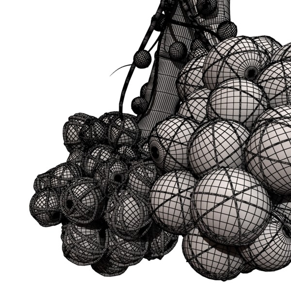
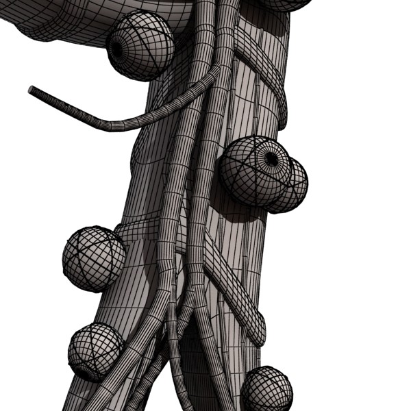
363.538 polygons. no smooth
436.828 polygons turbosmooth.
*******************************************
Detailed enough for close-up renders.
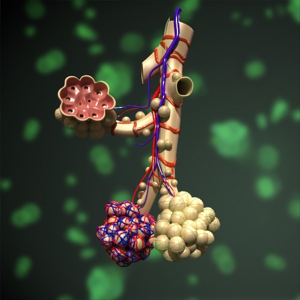
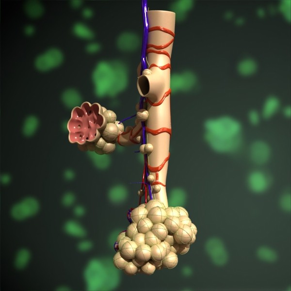
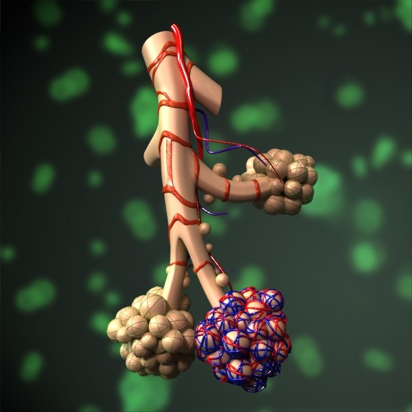
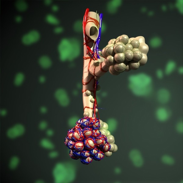
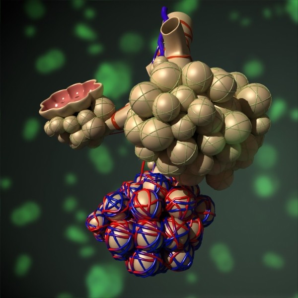
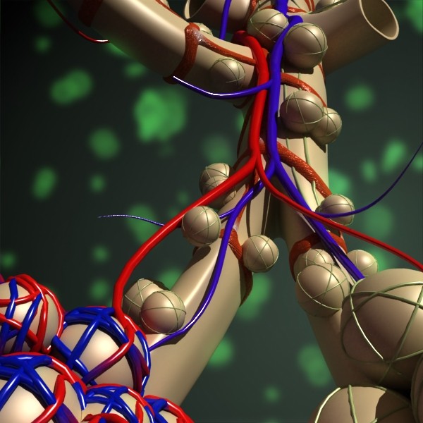
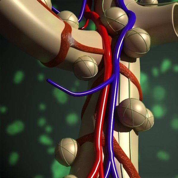
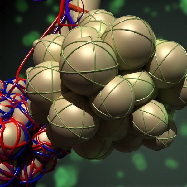
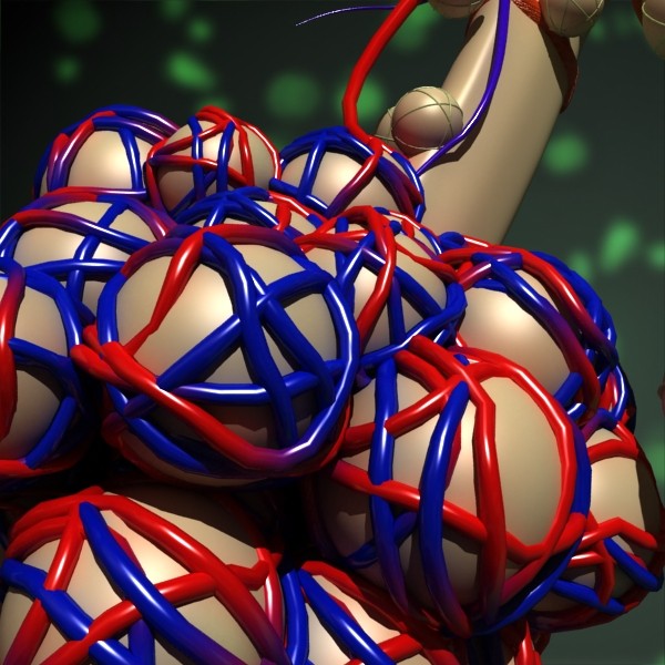
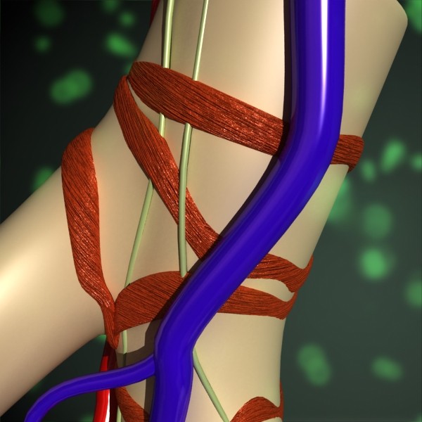
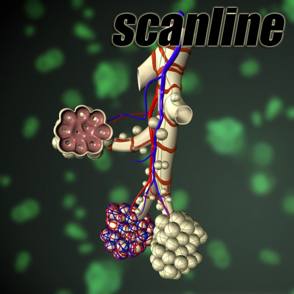
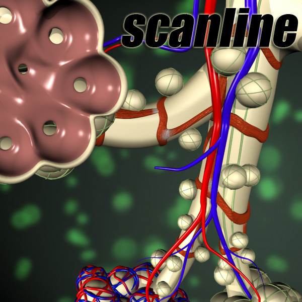
In the 1st order respiratory bronchioles, the number of alveoli in the wall is small, in the 2nd respiratory bronchioles there are 2 more, the 3rd order respiratory bronchioles are, even more, the alveolar passages are almost 100% formed by the alveoli, the alveolar sacs are completely formed by the alveoli. Alveolar sacs have a polygonal shape, separated by interalveolar septa 2-8 microns thick. The interalveolar septa are represented by the walls of the alveoli, the connective tissue elements located between them (elastic, collagen and reticular fibers) and the network of capillaries involved in gas exchange. Some alveoli communicate with each other due to holes in the interalveolar septa (“Kora pores”).
The total number of alveoli in both human lungs is 600–700 million. The diameter of one alveoli of a newborn child is on average 150 microns, an adult - 280 microns, in old age, reaches 300-350 microns. The total surface area of the alveoli varies from 40 m² when you exhale to 120 m²
The inner layer of the alveolar wall is formed by respiratory alveoli (alveoli of the 1st type) and large (secretory) alveoli (alveoli of the 2nd type), alveolar macrophages (alveoli of the 3rd type). Type 1 alveoli involved in gas exchange occupy a much larger area (97.5% of the inner surface of the alveoli) than type 2 alveoli (granular, cuboid, secretory cells). Like alveoli of the 1st type, alveoli of the 2nd type are located on the basement membrane; these cells produce a surfactant - a surfactant lining the inside of the alveoli and preventing them from falling. In the aggregate, alveoli of all types form the alveolar epithelium and lie on the basement membrane.
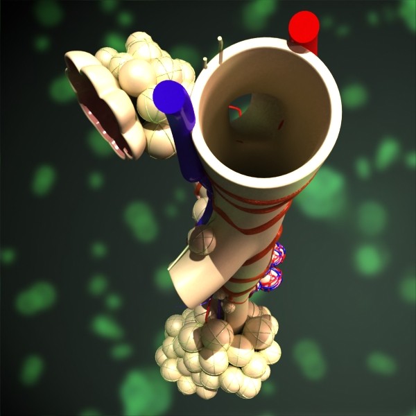
The aerohematic (air-blood) barrier between the alveolar air and the pulmonary capillaries is formed by the thinned cytoplasm of the respiratory alveoli, the basement membrane of the alveolar epithelium, and the wall of the pulmonary capillary and is 0.5 μm. In some places, the basement membranes diverge, forming cracks filled with elements of connective tissue. Each capillary is involved in gas exchange with several alveoli.
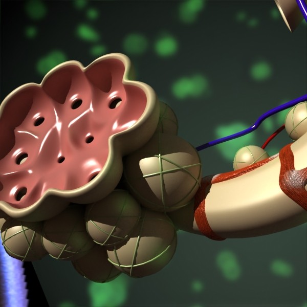
Alveoli are a hollow cup-shaped cavity found in the lung parenchyma where gas exchange takes place. Lung alveoli are found in the acini at the beginning of the respiratory zone. They are located sparsely on the respiratory bronchioles, line the walls of the alveolar ducts, and are more numerous in the blind-ended alveolar sacs.
Alveoli are particular to mammalian lungs. Different structures are involved in gas exchange in other vertebrates.
The alveoli consist of an epithelial layer of simple squamous epithelium, and an extracellular matrix surrounded by capillaries. The epithelial lining is a part of the alveolar membrane, also known as the respiratory membrane that allows the exchange of gases.
In the alveolar walls there are interconnecting air passages between the alveoli known as the pores of Kohn. The alveoli contain some collagen fibers and elastic fibers. The elastic fibers allow the alveoli to stretch when they fill with air during inhalation. Then they bounce back during exhalation in order to expel the carbon dioxide-rich air.
At FlatPyramid, we have created a 3D model of the Alveoli Anatomy. It is very well defined and with high-resolution. The model can be used for various purposes. It can be used in medical schools to demonstrate the students about its functioning and structure. It is available in different formats. We hope you like our model.
In case of any query, please feel free to contact us. We shall be glad to help you out.
Thank you for showing interest in the 3D model of the Alveoli Anatomy.