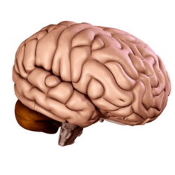
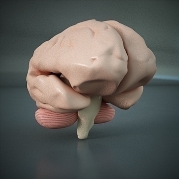
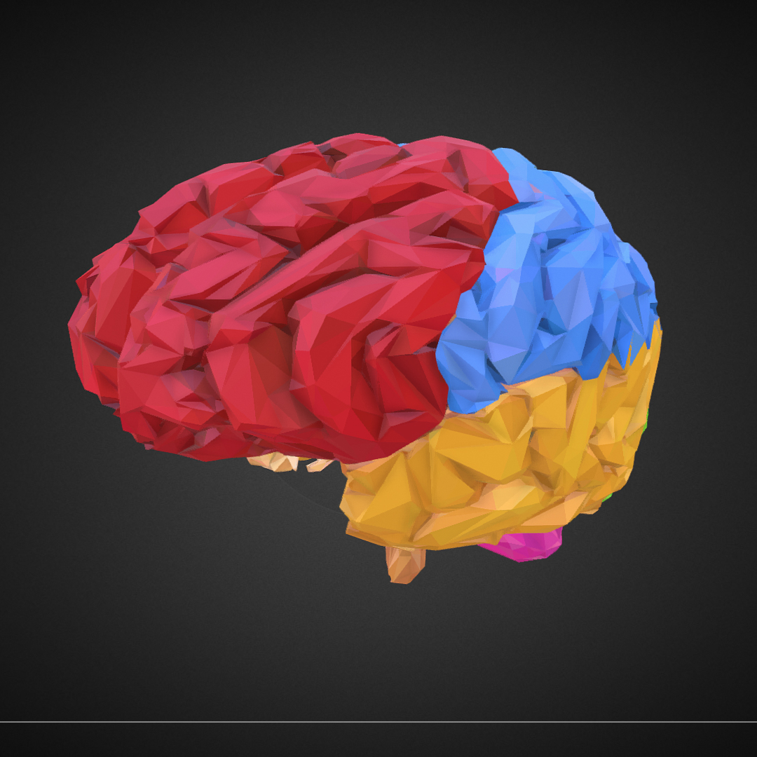
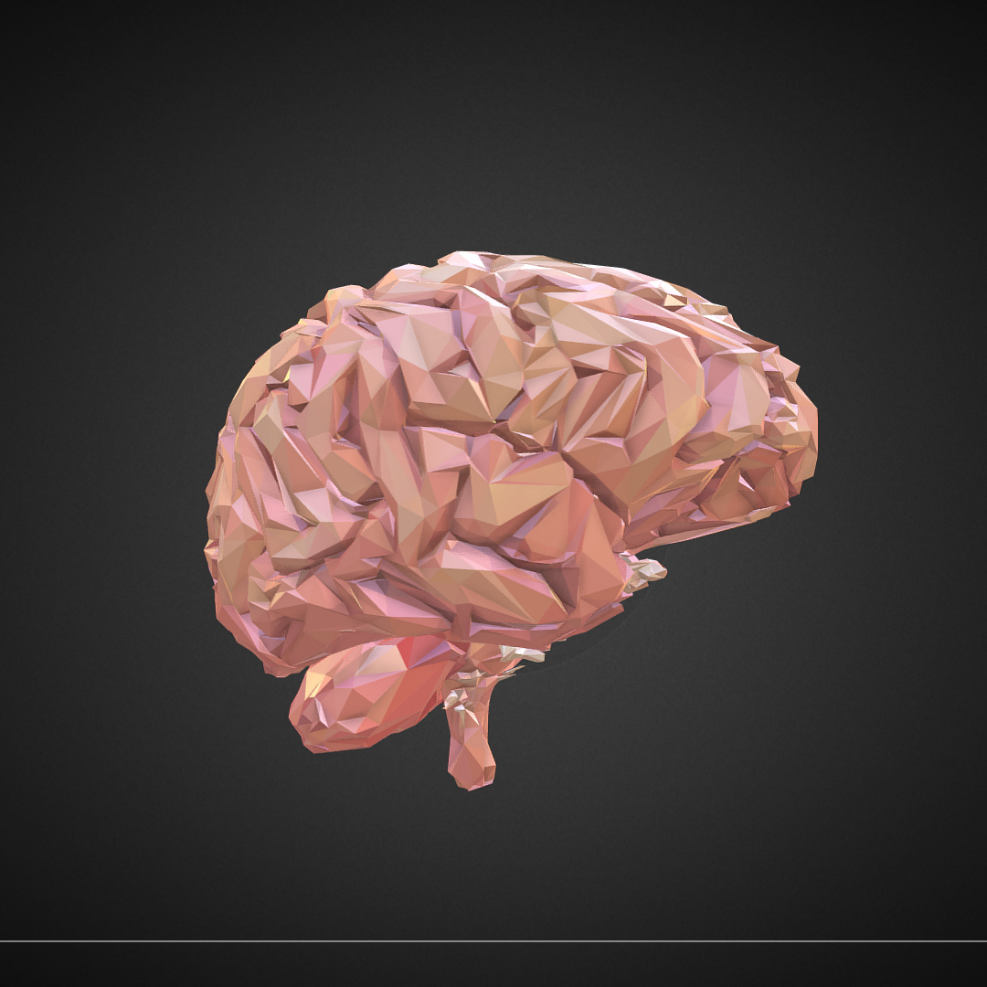
Brain 3D model free on Flatpyramid.
The brain is the central department of the central nervous system (neuraxis) of all vertebrates, in which it is contained in the "box" - the skull. Also, the brain is found in many invertebrates with different types of nervous system. The process of evolutionary formation of the brain is called "cephalization".The brain consists of different types of neurons that form the gray matter of the brain (bark and nucleus). Their processes (axons and dendrites) form a white matter. White and gray matter, as well as neuroglia, form a nervous tissue, from which, including the brain. Brain neurons communicate with each other and with the neurons of other parts of the nervous system due to universal nerve bonds - synapses.The structures
The structures of the human brain are responsible for fulfilling a variety of tasks: from the control of vital (vital) functions to higher mental activity.The development of the central nervous system (CNS) and the ganglia in the invertebrates has certain similarities to the vertebrates. First of all, the nervous system in them is an ectodermic derivative. Second, the central nervous system is formed as a result of the migration of neurons.The difference lies in the fact that the vertebrate ectoderm, from which the central nervous system appears, is located dorsally. Experiments on Drosophila and Caenorhabditis elegans have shown that the "nervous" ectoderm is located ventrally (Drosophila), or migrates from the lateral side to the anterior (C. elegans), and then immersed in the thickness of the embryo. The next stage is the formation of a "brain", that is, the conglomeration of neurons in the anterior ganglia.The nervous system of the vertebrates is a derivative of the nerve plate, and it, in turn, is also a derivative of the ectoderm. Subsequently, the nerve plate turns into a nerve tube. In the middle of the tube formed the same form of the cavity - neurocelum. It is in the cranial region of the nervous tube and the brain develops. However, it should be noted that cerebral thickening is still present in the nerve plate.The nerve tube consists of plates: ventral, dorsal and lateral. The lateral plate of its length is divided by the intermediate sulcus (sulcus Gisa) on the ventralnolateral (basal) and dorsolateral (auric (wing)) plates. These plates, with further development, are found in the spinal cord, oblong and median. From the basal plate will be formed motor components, from the alkaline - sensitive.Stages of brain development
The first stage in the development of the brain is the appearance of the anterior fold of the brain (Latin plica ventralis encephali). She divides the existing thickening into two "regions": archencephalon, which is located in front of the mitochondria and deuteroencephalon, which is located behind it.The next stage of development is the stage of the three primary bladders: the prostate brain (Latin prosencephalon), the median brain (mesencephalon), and the rhombencephalon (Latin rhombencephalon).The first bubble is an archencephalon derivative, the other two are deuteroencephalon. The stage of the three bubbles goes into the stage of five tertiary: the anterior brain is divided into the ultimate brain (Latin telencephalon) and the intermediate brain (Latin diencephalon); The rhomboid brain is divided into the back (Latin metencephalon) and oblong (Latin myelencephalon seu medulla oblongata).The middle brain is not shared. Subsequently, the posterior brain gives rise to a cerebellum and a bridge (the latter develops only in mammals). During development, one part of the brain grows faster than others, which causes (in reptiles, birds, and mammals) brain arches: cerebral, bridge (only in mammals and cervical).The neurocele of the rhomboid brain is converted into a fourth ventricle, the middle one - in the water supply (Latin aqueductus), the intermediate - in the third ventricle and the terminal - in the first and second ventricles.Download Brain 3D model free on Flatpyramid now.